Organizado por el IRB Barcelona en colaboración con la Fundació Catalunya – La Pedrera.
Los tutores de esta edición son: Nareg Djabrayan, Julia García, Saska Ivanova, Anabel-Lise Le Roux, Berta Terré, Natalia Trempolec, Montse Fàbrega, Joana Raquel Faria, Sylwia Gawrzak, Ivan Ivani, Samira Jaeger y Rosa María Ramírez.
Presentation
Este vídeo ha sido preparado por los estudiantes del programa Locos por la Biomedicina 2014.
Objectives
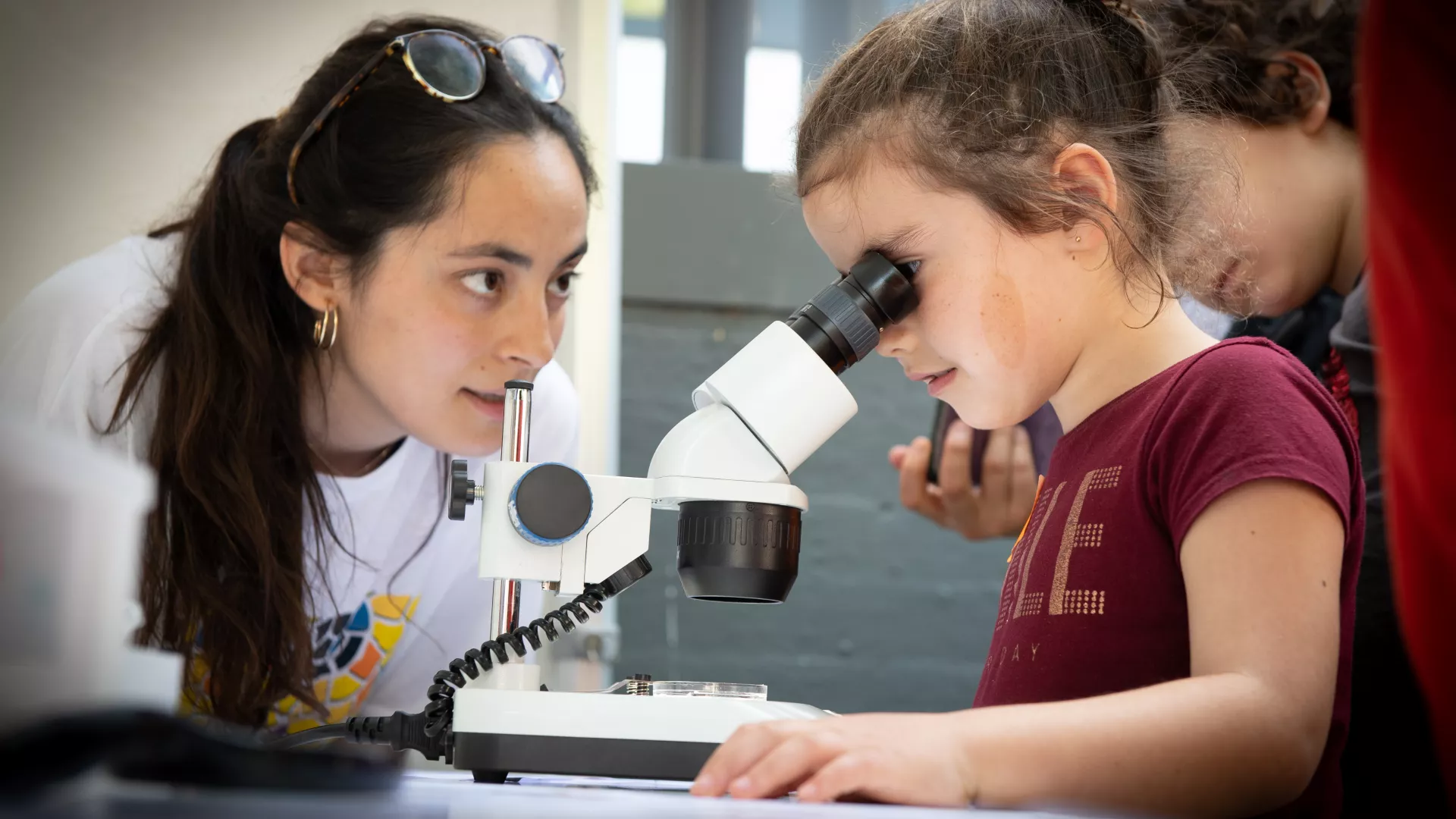
Crazy About Biomedicine is a year-long course directed at students in their first year of baccalaureate, who wish to explore some of the exciting discoveries currently being made in the life sciences. Through this course, students will have a chance to deepen their knowledge of scientific theory and techniques in the field of biomedicine. They will work alongside our young researchers to get a taste for what doing science in a top international research institute is like, gain some hands-on experience in the latest cutting-edge methodologies, and position themselves for a potential career in the life sciences.
Course Description
This course includes a mixture of theoretical lecture sessions and practical hand-on experimental activities, to take place on 18 Saturdays throughout the calendar year. The course will cover 12 current scientific topics, ranging from cell and molecular biology, to structural and computational biology and chemistry, presented by IRB Barcelona PhD students. In the first ‘semester’ (Jan-June 2014), the first 3 Saturdays will be dedicated to these general lectures for all participants. During the following 6 Saturdays, small groups will enter the labs for the hands-on practical sessions. This schedule will then be repeated with 6 new research topics for the second semester (June-Dec). Participating students must commit to attending for the duration of the year.
Course language
All talks and practical sessions will be conducted in English.
Course dates and times
The course will run from January to December 2014, 10.00-14.00h.
SEMESTER I
- Sat, 11 January 2014: Inauguration at La Pedrera
- Sat, 18 January 2014: Talks 1
- Sat, 1 February 2014: Talks 2
- Sat, 15 February 2014: Talks 3
- Sat, 22 February 2014: Practicals 1
- Sat, 8 March 2014: Practicals 2
- Sat, 15 March 2014: Practicals 3
- Sat, 29 March 2014: Practicals 6
- Sat, 5 April 2014: Practicals 4
- Sat, 26 April 2014: Practicals 5
SEMESTER II
- Sat, 10 May 2014: Talks 1
- Sat, 24 May 2014: Talks 2
- Sat, 7 June 2014: Talks 3
- Sat, 20 September 2014: Practicals 1
- Sat, 4 October 2014: Practicals 2
- Sat, 18 October 2014: Practicals 3
- Sat, 8 November 2014: Practicals 4
- Sat, 22 November 2014: Practicals 5
- Sat, 13 December 2014: Practicals 6
- Sat, 11 January 2015: Closing ceremony
Course fees
This course is free of charge, and includes access to the IRB Barcelona facilities and course materials. Lunches and snacks are not provided. Students will receive a Certificate of Participation upon successful completion of the course at a special ceremony on January 11, 2014, to which parents and teachers will be invited.
Course location
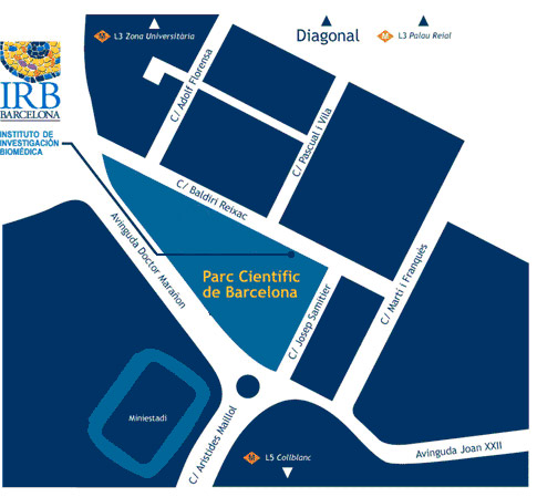
Institute for Research in Biomedicine (IRB Barcelona)
c/ Baldiri Reixac, 10
Barcelona
Who can apply
This course is directed toward students in the first year of their baccalaureate, who have a special interest and talent in the fields related to the life sciences (primarily biology and chemistry).
Students may apply to a maximum of 2 programmes within the "Crazy About Science" series, and can participate in only one.
How to apply
Interested students must fill in the online application form and include a letter of motivation. A letter of recommendation will be requested directly from two of their teachers, who should know the applicant well. If the applicant has recently changed school then the letters of recommendation should be requested from his/her old teachers. The deadline for registration is 17 October, 2013.
The course is open to a total of 24 students. Candidates will be selected on the basis of their academic record, teacher recommendations and motivation to participate. A short list of candidates will be invited for interviews with the scientific organizers in November after which the final selection will be made. Students will be informed of the outcome by the first week in December. The students selected to participate and their parents/guardians will be asked to sign a letter of commitment to attend all sessions.
Important dates
- Thurs, 17 October 2013: Application deadline
- Week of 28 October 2013: Short-listed candidates contacted
- 18-29 November 2013: Interviews
- 1-5 December 2013: List of selected students published
- Sat, 11 January 2014: Inauguration at La Pedrera
- Sat, 18 January 2014: Course begins
Collaborators
Fundació Catalunya La Pedrera, Parc Científic de Barcelona


If you have any question, please contact us at: irb_outreach@irbbarcelona.org
Programme
SEMESTER 1
1. Prelude to metamorphosis: Cells in transition
Nareg Djabrayan (Milán Lab)
Metamorphosis is defined as the abrupt, developmentally programmed change in the form of an animal. This change in structure involves the remodeling and even complete replacement of juvenile organs with those of the adult. Aside from the curious notion of an “animal within an animal”, the phenomenon of metamorphosis provides the opportunity to study many of the cellular behaviors associated with stem cells and cancer. These include the specification and maintenance of stem cell populations, the controlled transition from quiescence to proliferation, and the eventual changes in cellular architecture that allow for the remodeling of larval organs to their final adult forms.
The larva of Drosophila melanogaster undergoes a complete transformation from a vermiform “maggot” with no limbs to a winged six-legged fly. During the transition from the larval to adult form, most larval tissues are destroyed and replaced by organs arising from populations of “imaginal” cells. The adult trachea arises from a population of imaginal tracheal cells which for most of larval life remain quiescent. Before metamorphosis begins, imaginal tracheal cells proliferate and change their cellular architecture in preparation for the remodeling that must take place during metamorphosis.
This section will focus on the changes in cell behavior of imaginal tracheal cells leading up to the larval to adult transition. In the practical course, students will utilize genetic and cell biological tools to observe this process and interrogate the underlying mechanisms that ensure the specification, maintenance and activation of imaginal tracheal cells.
2. Designing peptide drugs
Júlia Garcia (Giralt Lab)
Peptides are small proteins made up of a small number of amino acids. The distinction between proteins and peptides is somewhat arbitrary, although proteins typically contain 50 or more amino acid, whereas peptides contain fewer.
Natural peptides are present in our organism where they play several biological roles. biological roles. The main ones involve interaction with macromolecules, such as proteins. For this reason, peptides can be used as therapeutic agents in order to treat different diseases. These potential drugs can be chemically synthesized by taking advantage of a method called solid-phase peptide synthesis (SPPS). While this technique offers numerous advantages, it is crucial that the method is designed properly in each case.
Since the most biological targets are usually found inside cells, peptides need to be able to cross cell membranes, which act as barriers. Certain types of peptides, such as some cyclic or bicyclic ones, have proved, have proved to possess this ability when some specific elements are introduced into their sequences. In the practical session, we will attempt to design and synthesize a therapeutic peptide.
3. Autophagy: cellular self-eating mechanism
Saska Ivanova (Zorzano Lab)
Every day cells have to deal with many types of damage and assaults, and these must be resolved in order to ensure that cells function properly and stay healthy. Unresolved damage normally leads to cell death. One of the main cellular “defense” mechanisms is autophagy, a process which, in basal conditions, is responsible for protein turnover and organelle quality control. However, under conditions of stress, autophagy is induced and functions as a pro-survival mechanism, clearing accumulating protein aggregates and/or damaged organelles. Additionally, in starvation, autophagy replenishes nutrients by breaking down cellular components. In the last decade, autophagy imbalance (up- or down-regulation) has been linked to many diseases, including diabetes, cancer, and neurodegeneration. In this regard, the mechanisms and signaling pathways involved in autophagy are extensively studied in order to gain deeper knowledge into how this process can be regulated and possibly manipulated in a disease-specific manner.
Delving into the main autophagy pathways, in this course we will focus on how to induce and biochemically recognize autophagy. The students will work directly with cells and gain insight into the main techniques used in autophagy and cellular biology.
4. The role of biological membranes in cells
Anabel-Lise Le Roux (Pons lab)
Cells and organelles are surrounded by membranes, which separate, organize and act as scaffolds for many processes. Membranes are formed by a “lipid bilayer” – a two-layered structure composed of insoluble phospholipids molecules that act as barrier to keep other molecules in the cell where they are needed and prevent them from traveling into areas where they are not. Phospholipids can also form other sub-cellular structures, such as “vesicles”, balloon like structures that transport their contents throughout the cell.
Fine-tuning of the chemical and physical properties of biological membranes give them their crucial role in the cell. How? What are exactly these phospholipid molecules and how do they form this barrier that is so resistant and flexible at a time? How can this barrier be crossed to enable communication of the cell with its external environment? What prevents the cell from exploding? Crucial partners in the functions of biological membranes are proteins, actually present in huge amount in the bilayer. What are the different types and roles of membrane proteins, what is the interplay between proteins and the phospholipid bilayer? How are proteins moving in the bilayer and how do they organize to fulfill their functions?
In the practical session, we will study a membrane protein that is an oncogene if overexpressed or deregulated. Part of its regulation is due to membrane association and interaction. We will study the affinity of the protein towards lipid bilayers of different compositions using a lipid sedimentation assay.
5. Understanding DNA: What makes you different from everybody else?
Berta Terré (Stracker Lab)
It can be said that DNA is the genetic “instruction manual” found in all our cells. This molecule contains genetic information that control several cellular functions such as proliferation, apoptosis and even the color of our eyes. However, sometimes the DNA can suffer some modifications that can disrupt all these biological processes leading to the development of several diseases such as cancer. Because of that, our cells have developed a system to control and repair these lesions: the DNA Damage Response pathway. Simply put, we have some genes that function as guardians of the genome, which are responsible for maintaining genomic stability and protecting our cells. However, this system does not work exactly the same way for everyone, and that is one of the reasons why despite having the same lifestyle, some people develop cancer while others do not.
In this two-part course, we will explore the association between genomic instability and cancer. We will try to understand the mechanisms by which DNA controls cellular function and how the genetic differences that exist between people can influence their risk of getting cancer.
This course will be continued in Semester II.
6. The role of p38 MAPK cascade in cancer development
Natalia Trempolec (Nebreda Lab)
Cells, in order to perform their functions and survive, have to be constantly aware of changes in their extracellular environment. In order to respond to large numbers of stimuli and environmental changes, they have developed sophisticated mechanisms that allow them to receive signals, transmit information and coordinate an appropriate cellular response. Since most extracellular particles are not able to enter the cell, signals must be passed into the cell. This signal transduction is based on intracellular “communication lines”, or signaling pathways, many of which operate when a series of chemical changes, called “phosphorylation”, takes place. Signal transduction involving phosphorylation is called a “protein kinase cascade”. The proper function of signaling pathways is crucial for cellular homeostasis and disruption of the balance results in disease, like cancer, diabetes or immune response inflammation.
Our laboratory studies a special protein kinase called p38 MAPK and its signaling cascade. It becomes activated in response to cellular stress, and plays a critical role in inflammation, cell cycle progression, differentiation, proliferation, cell growth and death. In this course, we will take a detailed look at the p38 MAPK signaling cascade, to understand when and how it is initiated, how it progresses and what happens to other molecules in the cell once p38 MAPK has been activated.
SEMESTER 2
1. Stepping into the world of proteins
Montse Fàbrega (Coll lab)
Proteins typically make up about half the total weight of the biomolecules in a cell. They play a wide variety of functional roles, including binding, catalysis, defence and signalling. Many studies involving proteins aim to achieve a detailed atomic structure of the protein in order to better understand its function, evolutionary development or interactions.
But working with proteins is a difficult and pain-staking process. First they have to be reproduced by a host cell to obtain enough quantity to be able to work with, and then isolated. Only once a researcher has successfully done these steps, can he or she then obtain images of its three-dimensional structure using the X-rays produced by a synchrotron.
In this course we will look at some of the current genetic engineering and structural biology tools and techniques that researchers use to determine the structures of proteins at the atomic level.
2. Understanding DNA: What makes you different from everybody else?
Joana Raquel Faria (Stracker Lab)
It can be said that DNA is the genetic “instruction manual” found in all our cells. This molecule contains genetic information that control several cellular functions such as proliferation, apoptosis and even the color of our eyes. However, sometimes the DNA can suffer some modifications that can disrupt all these biological processes leading to the development of several diseases such as cancer. Because of that, our cells have developed a system to control and repair these lesions: the DNA Damage Response pathway. Simply put, we have some genes that function as guardians of the genome, which are responsible for maintaining genomic stability and protecting our cells. However, this system does not work exactly the same way for everyone, and that is one of the reasons why despite having the same lifestyle, some people develop cancer while others do not.
In this two-part course, we will explore the association between genomic instability and cancer. We will try to understand the mechanisms by which DNA controls cellular function and how the genetic differences that exist between people can influence their risk of getting cancer.
This course is a continuation from Semester I.
3. Overview of metastasis: how do cancer cells colonize distant organs?
Sylwia Gawrzak (Gomis Lab)
Metastasis is a complex multistep process in which cancer cells leave the original tumor site and migrate to other parts of the body mainly through the bloodstream. Metastatic relapse, a late event in cancer progression, is associated with poor prognosis and is heavily implicated in the lethality of cancer. Although strategies targeting primary tumors have been improved, systemic treatments to prevent metastasis are less effective. Consequently, metastasis remains a challenging clinical problem, and the dissection of its biological mechanisms is crucial in order to reveal therapeutic targets to prevent and cure malignant tumors.
This course will give general overview of cancer biology and will focus on the different aspects of metastasis. We will learn about where cancer cells usually form metastases, the essential steps in this process, and the time it takes. The theoretical part will be illustrated with several examples of experimental techniques that are used to study metastasis. During the practical part, we will perform histological staining and compare several mouse tissue samples in order to identify metastatic lesions in various organs.
4. Computational drug discovery for HIV
Ivan Ivani (Orozco lab)
Drugs that attack HIV-1 protease, a protein related to AIDS syndrome, are one of the triumphs of modern medicine. This example demonstrates the power of structure-based drug design (SBDD): the atomic details of the viral protease structure fueled the successful design and development of five marketed drugs. Nowadays, to speed up and lower the costs of the SBDD, the use of computational tools is fundamental in the early stages of the process.
In this small project we will learn how the knowledge of protein structure can be combined with computational tools such as molecular docking to discover a new drug. We will study the case of HIV-1 protease, starting from basics about protein structure, the features of its binding pocket and later understanding how protein-ligand interactions can be evaluated.
Each student will visualize and analyze the features of our target protein structure and evaluate a compound using docking software. At the end of these steps we will be able to discriminate which compounds can be effective drugs.
5. Introduction to bioinformatics and systems biology
Samira Jaeger (Aloy Lab)
Bioinformatics has developed as an essential tool in various areas of biology. In molecular biology, different bioinformatics techniques are used to process and analyze large amounts of raw data. In other fields, such as genomics, bioinformatics aids in sequencing genomes, which yields important information for identifying disease-causing mutations.
The theoretical course will start with an introduction to bioinformatics and its significance for molecular biology and biomedicine. Later we will focus on an important field of bioinformatics, that is, systems biology, which looks at biological data at a more integrative level by means of biological networks. First, we will learn how such networks can be built and visualized using public databases. Next, we will study different computational methods that can be applied to analyze biological networks, including some that were originally developed for social networks and search engines. Last, we will discuss how outcomes of these analyses can be used in biomedicine.
In the practical “hands-on” session we will get to know the typical steps involved in bioinformatics analysis. We will learn how to use and combine computational tools to support biomedical research.
6. The skeleton of the cell
Rosa María Ramírez (Lüders Lab)
The cytoskeleton is a macromolecular network in eucaryotic cells that provides the structure and shape of cells, mechanical strength, locomotion, intracellular transport of organelles, and chromosome separation in mitosis and meiosis. The cytoskeleton is made up of diferent proteins that associate to form arrays of protein fibers, including actin filaments, intermedia filaments and the microtubules. These are long tubes that extend throughout the cell, providing an organizational framework for organelles and supporting cell movement. In mitosis the microtubules grow from the centrosomes to toward the chromosomes forming the mitotic spindle, which plays an important role in cell division. Cancer cells often have abnormal centrosomes and defects in the spindles. Microtubules are therefore an important target for cancer research.
In this course, we will learn about the microtubular cytoskeleton, its role in supporting cell dynamics, and its implications in health disorders. In the practical sessions we will analyze microtubule cytoskeleton organization during the cell cycle, using tissue culture cells and fluorescent light microscopy techniques.
Venue
CRAZY ABOUT BIOMEDICINE will take place at the IRB Barcelona facilities.

Venue
IRB Barcelona
c/o Parc Cientific de Barcelona
Carrer Baldiri Reixac, 10
08028 Barcelona
(Campus de la Diagonal, Universitat de Barcelona)

