Organizado por el IRB Barcelona en colaboración con la Fundació Catalunya – La Pedrera.
Los tutores de esta edición son: Francisco Freixo, Helena González, Benjamí Oller, Bahareh Eftekharzdeh, Anabel-Lise Le Roux, Michela Candotti, Jordina Guillen, Ana Ferreira, Julia García, Sabine Klischies, Natàlia Trempolec, Juliana Amodio y Montse Fàbrega.
Presentation

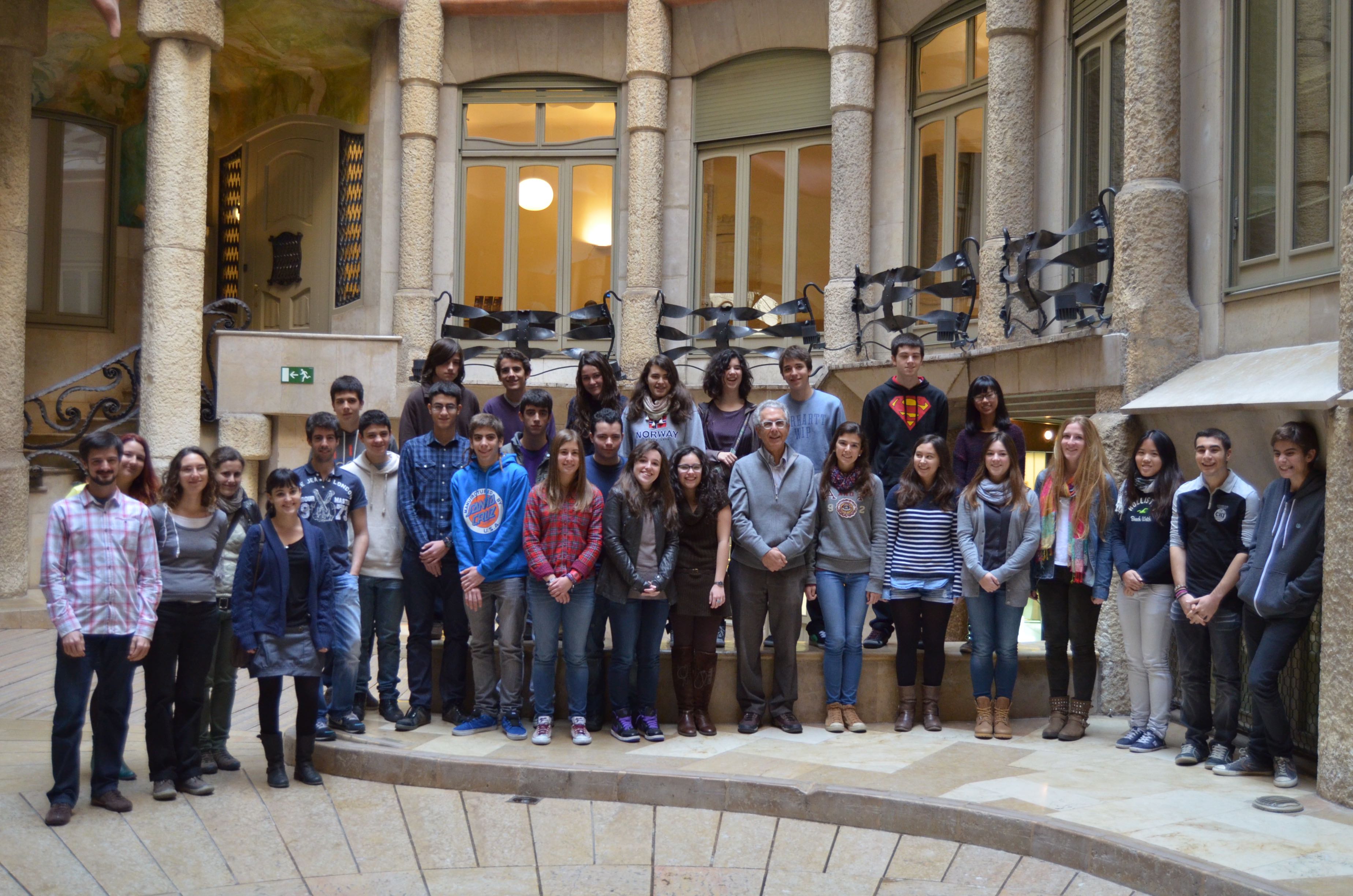
Participantes del curso “Crazy About Biomedicine”, edición 2013.
Objectives
Crazy About Biomedicine is a year-long course directed at students in their first year of baccalaureate, who wish to explore some of the exciting discoveries currently being made in the life sciences. Through this course, students will have a chance to deepen their knowledge of scientific theory and techniques in the field of biomedicine. They will work alongside our young researchers to get a taste for what doing science in a top international research institute is like, gain some hands-on experience in the latest cutting-edge methodologies, and position themselves for a potential career in the life sciences.
Course Description
This course includes a mixture of theoretical lecture sessions and practical hand-on experimental activities, to take place on 18 Saturdays throughout the calendar year. The course will cover 12 current scientific topics, ranging from cell and molecular biology, to structural and computational biology and chemistry, presented by IRB Barcelona PhD students. In the first ‘semester’ (Jan-June 2013), the first 3 Saturdays will be dedicated to these general lectures for all participants. During the following 6 Saturdays, small groups will enter the labs for the hands-on practical sessions. This schedule will then be repeated with 6 new research topics for the second semester (June-Dec). Participating students must commit to attending for the duration of the year.
Course language
All talks and practical sessions will be conducted in English.
Course dates and times
The course will run from January to December 2013, 10.00-14.00h.
SEMESTER I
- Sat, 12 January 2013: Talks 1
- Sat, 26 January 2013: Talks 2
- Sat, 9 February 2013: Talks 3
- Sat, 23 February 2013: Practicals 1
- Sat, 2 March 2013: Practicals 2
- Sat, 16 March 2013: Practicals 3
- Sat, 6 April 2013: Practicals 4
- Sat, 20 April 2013: Practicals 5
- Sat, 4 May 2013: Practicals 6
SEMESTER II
- Sat, 18 May 2013: Talks 1
- Sat, 1 June 2013: Talks 2
- Sat, 15 June 2013: Talks 3
- Sat, 21 September 2013: Practicals 1
- Sat, 5 October 2013: Practicals 2
- Sat, 19 October 2013: Practicals 3
- Sat, 9 November 2013: Practicals 4
- Sat, 23 November 2013: Practicals 5
- Sat, 14 December 2013: Practicals 6
- Sat, 11 January 2014: Closing ceremony
Course fees
This course is free of charge, and includes access to the IRB Barcelona facilities and course materials. Lunches and snacks are not provided. Students will receive a Certificate of Participation upon successful completion of the course at a special ceremony on January 11, 2014, to which parents and teachers will be invited.
Course location
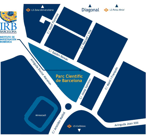
Institute for Research in Biomedicine (IRB Barcelona)
c/ Baldiri Reixac, 10
Barcelona
Who can apply
This course is directed toward students in the first year of their baccalaureate, who have a special interest and talent in the fields related to the life sciences (primarily biology and chemistry).
How to apply
Interested students must fill in the online application form and include a letter of motivation. A letter of recommendation will be requested directly from two of their teachers. The deadline for registration is October 31, 2012.
The course is open to a total of 24 students. Candidates will be selected on the basis of their academic record, teacher recommendations and motivation to participate. A short list of candidates will be invited for interviews with the scientific organizers in November after which the final selection will be made. Students will be informed of the outcome by the first week in December. The students selected to participate and their parents/guardians will be asked to sign a letter of commitment to attend all sessions.
Important dates
- Wed, 31 October 2012: Application deadline
- Week of Mon, 5 November 2012: Short-listed candidates contacted
- Weeks of November 12/19: Interviews
- Mon, 26 November 2012: List of accepted students published
- Sat, 12 January 2013: Course begins
Collaborators
Fundació Catalunya La Pedrera, Parc Científic de Barcelona


Programme
SEMESTER 1
1. Architecture of the cell – PART ONE
Francisco Freixo (Lüders Lab)
The cells of all living beings have a “skeleton” that provides support for all their functions, such as the transport of proteins throughout the cell, organization of the cell shape, and even cell motility, migration, and division. In human cells and other eukaryotic cells, this “cytoskeleton” is made up of a number of structures, including “microtubules”, which are tiny cylinders that can shrink, elongate, bundle, and distribute throughout the cytoplasm, according to the cell’s needs. Other important structures in the cytoskeleton include “centrosomes” and “mitotic spindles”, which play an important role in cell division, helping to ensure that the genetic material of a dividing cell will be equally split into each of the two new cells. Centrosomes and mitotic spindles are often found to be abnormal in cancer cells, and therefore these structures are of great interest in cancer research.
In this two-part course, which will run over both semesters, we will learn about the components of the cytoskeleton, and take a look at why they are so important for the cell. In the practical exercises, we will show you how microtubules organize inside a cell (in arrays of various types and shapes), and how the cells depend on these arrays. We will analyze the molecules that regulate where and when different cell types form their microtubules, using tissue culture cells and mouse hippocampal neurons, and fluorescent light microscopy techniques.
This course will be followed by a second session in Semester II, led by Sabine Klischies
2. What does DNA damage have to do with cancer?
Helena González (Stracker Lab)
DNA is a long molecule that contains “genetic information”, a coded instruction manual for the cells. Everything the cells do is somehow coded in DNA. It determines, for example, which cells should grow and when, which cells should die and when, which cells should make hair and what color it should be. Simply put, DNA tells cells how they should behave correctly.
But sometimes the DNA becomes “mutant”, which means it has been damaged or broken. As a result, these instructions can be misread, leading to the development of diseases such as cancer. Fortunately cells have a very effective system to repair these lesions: the DNA Damage Response pathway. Its job is particularly difficult considering that DNA is constantly attacked by toxic agents (like sun radiation, pollution, tobacco). Its function is extremely important because a failure to repair DNA lesions may result in cancer.
This course will give important clues to understand the link between the factors that cause DNA damage and the development of cancer. We will also have the opportunity see what happens inside the nucleus of a cell after it has been damaged. Can it be repaired?
3. Peptides as shuttles to carry antibodies across the BBB
Benjamí Oller (Giralt Lab)
Peptides are small proteins, which are biomolecules made out of amino acids. One of their main roles is to interact with other molecules, generally bigger proteins, in order to initiate or prevent their function. Some peptides in particular are recognised by protein receptors on cell membranes and are internalized. This natural function can be used in some cases to transport bioactive compounds that are unable to cross certain barriers.
One of the most impenetrable biological barriers is the one that separates the blood from the brain, the blood-brain barrier (BBB). This structure is the main hurdle for drugs meant to treat illnesses affecting the central nervous system, such as Alzheimer’s and Parkinson’s diseases or brain cancers. Some potentially effective treatments for brain diseases are based on proteins, but most of them cannot cross the BBB so they have a very local effect that is often not enough to overcome the illness. Linking peptides that are able to cross the BBB to these proteins can greatly improve their distribution and thus their efficacy. However, decorating protein drugs with peptides without affecting their activity is not an easy task. Proteins have many reactive groups on their surface and only some of them should be modified. Organic chemistry offers many different strategies to meet this challenge, which will be explored in this seminar. We will learn about reactions that allow us to modify the side chains of certain amino acid residues, the sugars that are often linked to them or the N-terminus of the molecule. Ultimately, in the practical session, we will perform a selective ligation and we will analyse the outcome of the reaction.
This course will be followed by a second session in Semester II where the concept of selective reactivity will be revisited in the chemical synthesis of peptides. (Designing peptide drugs, Julia García)
4. An introduction into neurodegeneration and neurodegenerative diseases
Bahareh Eftekharzdeh (Salvatella lab)
Alzheimer’s disease is the most common form of dementia. It has no cure, and symptoms worsen as the disease progresses, eventually leading to death. It was first described by German psychiatrist and neuropathologist, Alois Alzheimer in 1906. The causes and progression of Alzheimer's disease are not well understood. Research indicates that the disease is associated with plaques and tangles in the brain. Current treatments only help with the symptoms of the disease, but none stops or reverses its progression. As of 2012, more than 1000 clinical trials have been conducted to find ways to treat Alzheimer’s, but it is unknown whether any of the treatments being tested will work. Mental stimulation, exercise, and a balanced diet have been suggested as ways to delay cognitive symptoms (though not brain pathology) in healthy older individuals, but there is no conclusive evidence supporting an effect. This course will take an in-depth look at the mechanisms of neurodegeneration, with a focus on two main ones in Alzheimer’s and Parkinson’s disease.
We will begin with an introduction to the history of these diseases, looking at the first times they were detected in brains of patients. This will be followed by an in-depth explanation of the molecular and cellular mechanisms of disease. Next we will go through the hypotheses for the causes, and the latest research approaches to finding cures, including the latest medical strategies. Finally we will finish the course with a question and answer session where students will have the opportunity to think about the possibilities and approaches in this field of research for the future.
5. The role of lipid membranes in cells
Anabel-Lise Le Roux (Pons Lab)
Cells and organelles are surrounded by membranes, which separate, organize and act as scaffolds for many processes. Membranes are formed by a “lipid bilayer” – a two-layered structure composed of insoluble lipid (or fat) molecules that acts as barrier to keep other molecules in the cell where they are needed and prevent them from traveling into areas where they are not. Lipids can also form other sub-cellular structures, such as “vesicles”, small bubbles that transport their contents throughout the cell. A special type of vesicle called a “liposome” can be formed artificially and used as a vehicle to administer nutrients or drugs, therefore highlighting the important role that lipid membranes can play in the treatment of disease.
This course will begin with an introduction to the basic properties of biological membranes: Why are they insoluble in water? Why can detergents dissolve lipids in water? What is the role of vesicles? Why is membrane fusion useful? We will also take a closer look at the contribution of membranes to cell function. How can molecules cross the lipid bilayer? What prevents cells from exploding? The practical work will consist in producing some artificial lipid multilayers, in the shape of vesicles.
6. Computational drug discovery for HIV
Michela Candotti (Orozco Lab)
Drugs that attack HIV-1 protease, a protein related to AIDS syndrome, are one of the triumphs of modern medicine. This example demonstrates the power of structure-based drug design (SBDD): the atomic details of the viral protease structure fueled the successful design and development of five marketed drugs. Nowadays, to speed up and lower the costs of the SBDD, the use of computational tools is fundamental in the early stages of the process.
In this small project we will learn how the knowledge of protein structure can be combined with computational tools such as molecular docking to discover a new drug. We will study the case of HIV-1 protease, starting from basics about protein structure, the features of its binding pocket and later understanding how protein-ligand interactions can be evaluated. Each student will visualise and analyse the features of our target protein structure and evaluate a compound using docking software. At the end of these steps we will be able to discriminate which compounds can be effective drugs.
SEMESTER 2
1. Taking a look at frog oocytes
Jordina Guillen (Méndez lab)
This course will give a general overview of how to use the African clawed frog, Xenopus laevis, as an animal model system. In our laboratory we use Xenopus laevis oocytes to study the meiotic cell cycle and the translational control of maternal messenger RNAs (mRNAs).
Meiosis is a type of cell division that reduces the number of chromosomes in the parent cell by half and produces gamete cells. During meiosis there is no transcription and the key protein activities that drive this process are mainly controlled at the level of translation of previously stored maternal mRNAs. A family of proteins called CPEB (cytoplasmic polyadenylation element binding protein) are responsible for the translational control of maternal mRNAs by a process called “cytoplasmic polyadenylation”.
Although we will focus on the meiotic cell cycle and the first embryonic divisions, gene expression regulation at the level of translation of mRNAs controls other biological processes such as cell proliferation, differentiation, senescence, synaptic plasticity and angiogenesis, as well as pathological processes such as tumor progression.
In the practical course we will do three different experiments with Xenopus laevis oocytes: 1) in vitro fertilization of frog eggs and observation of the first embryonic divisions. 2) Selection and micro-injection of oocytes. 3) Observation of oocyte chromosomes aligned in the second metaphase plate, under normal conditions and in the absence of an essential protein for meiosis (CPEB4).
2. Looking for the appropriate size: genetics under control
Ana Ferreira (Milán Lab)
As an animal develops, its body parts and organs must grow to achieve the proper size and shape that is characteristic of each species. This growth has to be coordinated between and within the different organs in order to generate fully functional adults. How organs achieve a particular size and pattern is one of the fundamental questions in developmental biology today.
Our laboratory studies the cellular and molecular mechanisms underlying the regulation of tissue growth during normal development. For this purpose, we use the fruit fly Drosophila melanogaster, a well-described animal model often used in developmental biology. The fruit fly has many advantages as a model system, including the fact that it is well suited for genetic and molecular studies. Specifically, we focus on the wing “imaginal disc”, which grows and proliferates during the larval stages of the fly to give rise to the adult wing. The size and shape of the final adult wing is determined by the rates of cell division, cell growth and cell death during wing disc development. To reach a proper size and shape tissue and cell growth must be carefully controlled at the molecular and genetic levels. Otherwise this can lead to uncontrolled growth and proliferation, characteristic of cancer cells.
During this course, we will take a look at imaginal disc development and see first-hand how effective Drosophila can be as a model system. We will use classic genetics and confocal microscopy techniques to observe how size can be affected at the level of the organ and organism.
3.Designing peptide drugs
Júlia Garcia (Giralt Lab)
Peptides are small proteins made up of a small number of amino acids. The distinction between proteins and peptides is somewhat arbitrary, although proteins typically contain 50 or more amino acid, whereas peptides contain fewer.
Natural peptides are present in our organism where they play several biological roles. biological roles. The main ones involve interaction with macromolecules, such as proteins. For this reason, peptides can be used as therapeutic agents in order to treat different diseases. These potential drugs can be chemically synthesized by taking advantage of a method called solid-phase peptide synthesis (SPPS). While this technique offers numerous advantages, it is crucial that the method is designed properly in each case.
Since the most biological targets are usually found inside cells, peptides need to be able to cross cell membranes, which act as barriers. Certain types of peptides, such as some cyclic or bicyclic ones, have proved, have proved to possess this ability when some specific elements are introduced into their sequences. In the practical session, we will attempt to design and synthesize a therapeutic peptide.
This course is a continuation of “Peptide shuttles” offered by Benjamí Oller in Semester I.
4. The Architecture of the cell - PART TWO
Sabine Klischies (Lüders lab)
The cells of all living beings have a “skeleton” that provides support for all their functions, such as the transport of proteins throughout the cell, organization of the cell shape, and even cell motility, migration, and division. In human cells and other eukaryotic cells, this “cytoskeleton” is made up of a number of structures, including “microtubules”, which are tiny cylinders that can shrink, elongate, bundle, and distribute throughout the cytoplasm, according to the cell’s needs. Other important structures in the cytoskeleton include “centrosomes” and “mitotic spindles”, which play an important role in cell division, helping to ensure that the genetic material of a dividing cell will be equally split into each of the two new cells. Centrosomes and mitotic spindles are often found to be abnormal in cancer cells, and therefore these structures are of great interest in cancer research.
In this two-part course, which will run over both semesters, we will learn about the components of the cytoskeleton, and take a look at why they are so important for the cell. In the practical exercises, we will show you how microtubules organize inside a cell (in arrays of various types and shapes), and how the cells depend on these arrays. We will analyze the molecules that regulate where and when different cell types form their microtubules, using tissue culture cells and mouse hippocampal neurons, and fluorescent light microscopy techniques.
This course is a continuation of the first session held in Semester I, led by Francisco Freixo.
5. The role of p38 MAPK cascade in cancer development
Natalia Trempolec (Nebreda Lab)
Cells, in order to perform their functions and survive, have to be constantly aware of changes in their extracellular environment. In order to respond to large numbers of stimuli and environmental changes, they have developed sophisticated mechanisms that allow them to receive signals, transmit information and coordinate an appropriate cellular response. Since most extracellular particles are not able to enter the cell, signals must be passed into the cell. This signal transduction is based on intracellular “communication lines”, or signaling pathways, many of which operate when a series of chemical changes, called “phosphorylation”, takes place. Signal transduction involving phosphorylation is called a “protein kinase cascade”. The proper function of signaling pathways is crucial for cellular homeostasis and disruption of the balance results in disease, like cancer, diabetes or immune response inflammation.
Our laboratory studies a special protein kinase called p38 MAPK and its signaling cascade. It becomes activated in response to cellular stress, and plays a critical role in inflammation, cell cycle progression, differentiation, proliferation, cell growth and death. In this course, we will take a detailed look at the p38 MAPK signaling cascade, to understand when and how it is initiated, how it progresses and what happens to other molecules in the cell once p38 MAPK has been activated.
6. Stepping into the world of proteins
Juliana Amodio/Montse Fabrega (Coll Lab)
Proteins typically make up about half the total weight of the biomolecules in a cell. They play a wide variety of functional roles, including binding, catalysis, defence and signalling. Many studies involving proteins aim to achieve a detailed atomic structure of the protein in order to better understand its function, evolutionary development or interactions.
But working with proteins is a difficult and pain-staking process. First they have to be reproduced by a host cell to obtain enough quantity to be able to work with, and then isolated. Only once a researcher has successfully done these steps, can he or she then obtain images of its three-dimensional structure using the X-rays produced by a synchrotron.
In this course we will look at some of the current genetic engineering and structural biology tools and techniques that researchers use to determine the structures of proteins at the atomic level.
Venue
CRAZY ABOUT BIOMEDICINE will take place at the IRB Barcelona facilities.

Venue
IRB Barcelona
c/o Parc Cientific de Barcelona
Carrer Baldiri Reixac, 10
08028 Barcelona
(Campus de la Diagonal, Universitat de Barcelona)

