Research information
Background
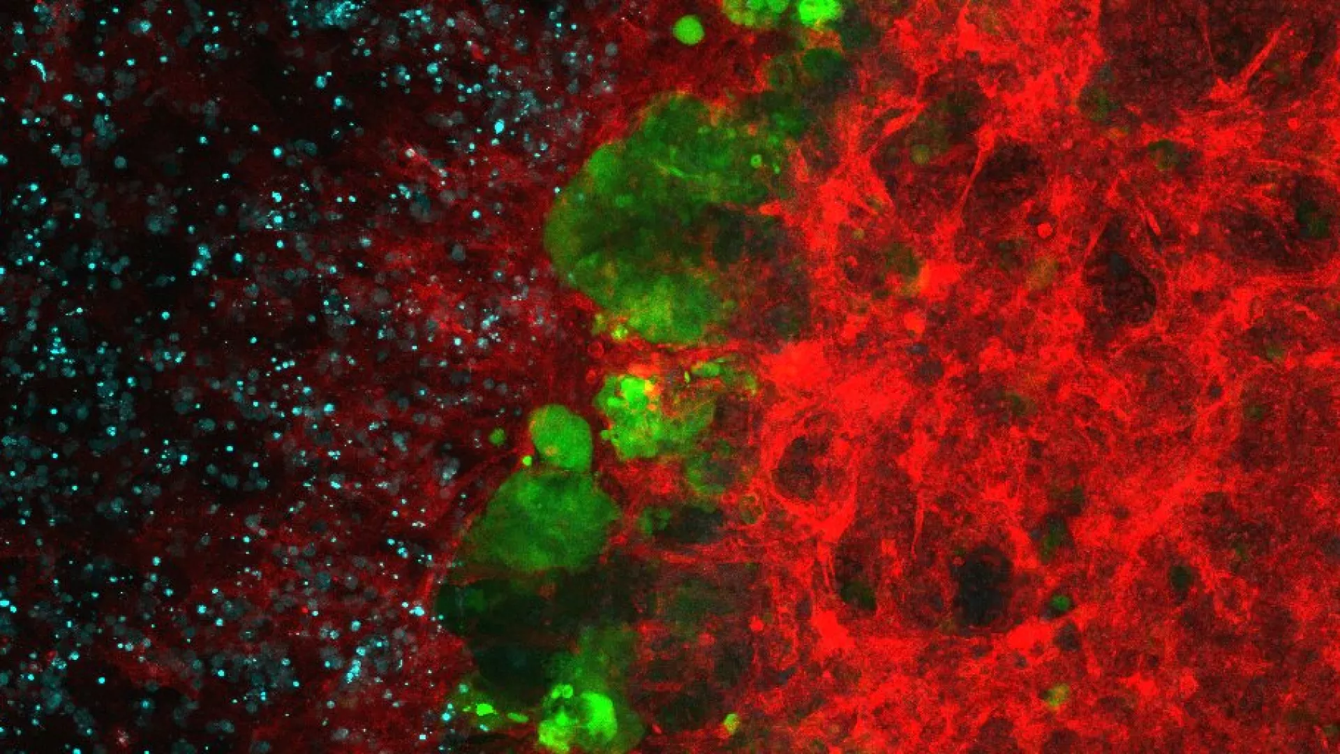
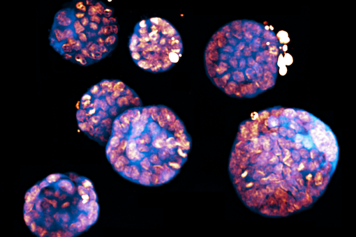
The Advanced Digital Microscopy Core Facility (ADM) was inaugurated in January 2009. The facility is committed to providing a complete range of advanced light microscopy imaging systems and techniques to researchers at IRB Barcelona and other institutions at the Barcelona Science Park (PCB), and external entities and visiting scientists. The facility provides full-time (24/7) access to state-of-the-art, mostly laser-based, commercial or customized microscopes, as well as assistance and development in Bioimage Analysis, data interpretation and presentation.
The ADM is also committed to develop research instrumentation to adapt, improve or newly implement innovative biology-driven imaging applications, for example in the field of mesoscopic imaging (light-sheet-based fluorescence microscopy).
The facility provides full-time 24/7 access to:
- Conventional transmission and epifluorescence microscopy and motorized fluorescence macroscopy
- Spectral confocal microscopy, including FRAP, FRET, Time-corelated Photon Counting (TCSPC) Fluorescence lifetime (FLIM), photoactivation and multiphoton microscopy
- Spinning disk confocal microscopy
- TIRF Microscopy
- High-content-Imaging automated microscopy equipped with epifluorescence
- 'High Content Screening' equipped with robotized loader and spinning disk
- Laser nanosurgery of living cells and organisms combined with FRAP
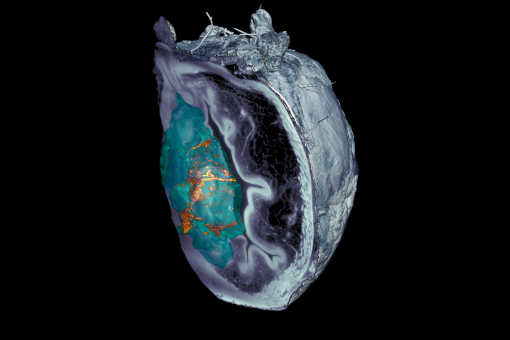
- Lightsheet based whole organ imaging with optical clearing
- Lightsheet-based SPIM for living organisms
- Image processing workstations, including a HIVE (Acquifer) storage with 240TB and computational units
- (from 2021) Lightsheet-based Oblique Plane Microscopy for large samples on inverted microscope.
For further details, please see our HOMEPAGE: https://adm.irbbarcelona.org/
For further information, contact us microscopy irbbarcelona.org
Selected publications
Projects
CERCAGINYS és la plataforma d’accés a les infraestructures científiques i tècniques dels 41 centres CERCA. La iniciativa vol optimitzar l’accés a aquestes instal·lacions per a tota la comunitat científica i tecnològica i, molt especialment, vol evidenciar la possibilitat que el sector privat empresarial i industrial també pot fer-ne ús, accedint, així mateix, als serveis de personal tècnic altament qualificat. La proposta forma part d’un projecte més ampli, vinculat al Pla d’Acció d’Infraestructures finançat pel Ministeri de Ciència i Innovació a través de la I-CERCA.

Services
Services for IRB Barcelona and external researchers (Public or Private)
- Spectral laser scanning confocal microscopy. 3D, 4D and 5D imaging with optical sectioning, custom spectral detection and resolution, multiple position or high-content mode, and incubated environment control for living cells.
Four LSM confocal units: Inverted confocal systems (Leica SP5, LSM 780, LSM880) and one upright confocal system (Leica SPE).
- Multiphoton microscopy. Tissue-deep fluorescence imaging by multiphoton excitations, and derived applications (SHG, etc…). Tailored on the SP5 with a MAITAI ultrashort pulsed IR laser (710-990nm).
- Spinning disk confocal microscopy. Fast 5D imaging with the Andor Revolution system, equipped with EMCCD camera technology, FRAP-Photoactivation, and microinjection.
Multimodal Super Resolution system for SIM/ISM/SMLM: The Zeiss Elyra PS1 system combines confocal microscopy (LSM880) with three modalities of super resolution: Image Scanning Microscopy (aka Airyscan), Structured Illumination Microscopy (SIM), and Single Molecule Localization Microscopy (SMLM: STORM, PALM and variants).
High Content Imaging/Screening with automated fluorescence widefield microscopy, confocal spinning disk or TIRF:
The Olympus ScanR enables fully automated inverted microscope for imaging wide areas, multiwell plates and multipositions, automated live imaging with temperature incubation and CO2 control, superstable illumination for reproducible and high content image analysis, and Total internal reflection microscopy (TIRF) for high contrast imaging at surfaces.
The Nikon LIPSI system enables fast High-Throughput Screening imaging with confocal spinning disk modality (CSU-W1 from Yokogawa) and multiple multiwell plates readouts, thanks to the integrated and multi-sample incubator that loads samples automatically. HTS can be performed on up to 20 plates. The system is also very suited to very long term imaging (1-2 weeks duration) by enabling the robot-driven loading of multiple experiments in parallel.Laser manipulation of living cells and organisms. FRAP and laser nanosurgery combined on a wide field fluorescence microscope to perform FRAP and fluorescence patterning, cell ablation, DNA damage, subcellular ablation and correlative microscopy.
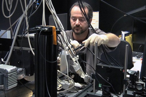
The Zeiss Axiovert 200M laser cutter is a custom platform equipped with a pulsed UV laser (355nm, 470ps) and spinning disk detection.Lighsheet Microscopy for optically cleared organs: A double sided macroscope-base detection and double-sided illumination lightsheet system for fluorescence and label free 3D imaging of tissues/organs/organisms ranging from 0.5mm to 2.5cm.
- Optical clearing: we offer expertise with the most recent optical tissue clearing techniques, including BABB (Murray’s clear), CUBIC, DISCO family, ECI, DeepClear, and more.
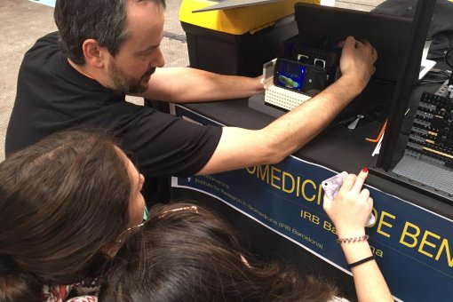
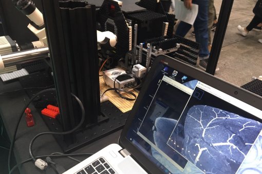
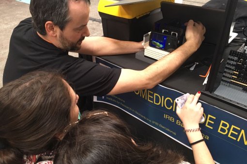
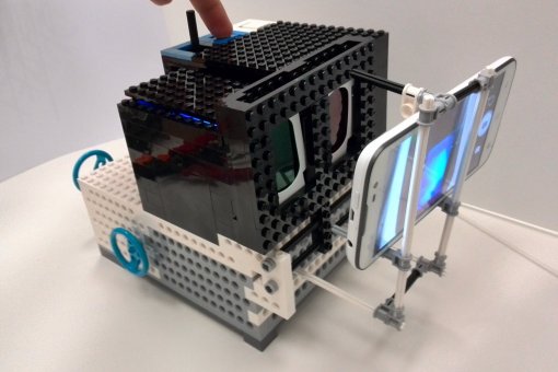
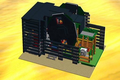
- Lightsheet Microscope based on LEGO bricks, LEMOLISH (http://legolish.org/lemolish) an open source instrument enabling lightsheet imaging of cleared organs of size up to 5cm.
- Lighsheet Microscopy for living organisms: A high-resolution double sided illumination SPIM system, including multiview and sample rotation, for full volume live imaging, for samples ranging from 50µm to 1mm.
- (From 2021) Lightsheet-based Oblique Plane Microscope: A custom built lightsheet based system on an inverted microscope to enable lightsheet imagingin multiwell plates and other conventional dish-like sample holders.
- (From 2021) High-throughput OPM. A highly automated OPM system for high-content imaging of organoids in multiwell plates, part of the MACH3CANCER (https://mach3cancer.org) project, funded by the AECC.
Bioimage Analysis and access to Imaging workstations: Image processing and analysis is available from three 64-bit workstations equipped with Imaris 9 (Bitplane), customized ImageJ/Fiji bundles, Matlab, CellProfiler, Voreen, Leica LAS AF, Igor Pro, and more. Custom Image Analysis workflows are developed on demand, tailored to imaging projects. Open source projects developed by ADM are also available, e.g. LOBSTER for large image data, MosaicExporerJ for large lightsheet datasets stitching, AutoscanJ for intelligent Microscopy, and more.
Conventional transmission and fluorescence microscopy. Bright field, phase contrast, differential interference contrast (DIC), dark field and spectral fluorescence imaging of fixed samples, color-based brightfield imaging (e.g. histological sections). Fluorescence stereoscopy/macroscopy for sample manipulation and dissection, extended focus imaging, live macroscopy.
Available instruments: Stereoscope, fluorescence Macroscope (Olympus MVX10), inverted epifluorescence microscope (ScanR).
Policies
Access
Our microscopy systems are available 24 hours a day, 7 days a week, to all scientists working in the Barcelona Science Park.
External scientists and companies can also access our facilities. Please send an request to access ADM services at microscopy irbbarcelona.org, describing the goal and principles of the experiments, the status of the project as per availability of probes/samples, and the target instrument(s) you would like to access. The facility will respond within 48h and, upon i] technical validation and ii] financial approval of our access terms, experiments planning will be organized according to availability and in the briefest delays.
Billing policies
We apply a 30% discount out of office hours (8am to 9am, 6pm to 10pm) and 50% discount for overnight imaging (from 10pm to 8am on confocal systems).
Booking
During office hours (9am-6pm), access to systems is restricted to a maximum of 4 hours per user. This restriction is released if booking is made less than 48h before the experiment. The High Content automated microscopy and Long-term Live imaging systems (ScanR and LIPSI) has no time booking restrictions.
Access to our booking application must be granted by the ADM staff for each new user and for each system, with the exception of the stereoscope, which is accessible to all. Booking is possible up to 14 days in advance.
In general, late cancellation (< 24h in advance) will result in billing if the time slot is not reserved by another user.
Some systems might have further booking restrictions (example: number of hours available per group per week). If you experience any problems or need special booking conditions for a specific experiment, please contact us.
To access the instrument booking application form, please click here. Please note that you will need to accept an SSL certificate when accessing the booking application.
